
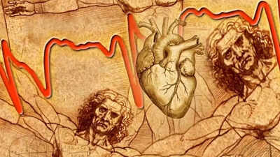
Leonardo da Vinci drew this heart structure 500 years ago: Scientists may have just solved a mystery that could predict heart disease
Leonardo da Vinci sketched the human body long before the advent of modern medicine. One of his more bizarre paintings focused on the inside of the human heart, which was barely understood by most people at the time. The 500-year-old building has long puzzled scientists, but they now think they know what Leonardo da Vinci was looking at. It’s a strange network of structures inside the heart called trabeculae.It’s been there in textbooks and scans for centuries, noticed but not really understood. Experts say it may even increase the risk of heart disease. It sounds a bit dramatic, but the science behind it is surprisingly solid, involving genetics, MRI scans and large-scale data from thousands of people. However, everything is not entirely clear yet. Some pieces of the puzzle are missing.
Leonardo da Vinci’s drawing of the heart and early discovery of trabeculae within the human heart
Leonardo da Vinci did not make blind guesses. He dissected human bodies himself, which was rare for his time and somewhat controversial. In his drawings of the heart, he noticed these branching, almost tree-like patterns within the ventricles. He thought they might warm the blood. Like a natural heating system. A creative idea. That’s not entirely true, experts say, but it’s not completely mentally abnormal either. For hundreds of years, these structures did not attract much attention. Sure, they’re anatomically visible, but mostly just ignored as internal texture. According to a study published in the journal Nature titled “Genetic and functional insights into the fractal structure of the heart‘, these structures are called trabeculae. They form a spongy, uneven lining inside the ventricles. More like tangled muscle bundles, they appear to be more than just residual biological noise from development. Researchers now believe they may actually affect blood flow and how efficiently the heart pumps blood. Some shapes appear to be associated with better heart function.
Large-scale MRI scan reveals trabecular patterns linked to heart disease risk
The scientists used MRI scans from a large-scale population study that included data from tens of thousands of people. One of the largest sources is UK Biobank. Some trabecular patterns appear to be associated with a higher risk of cardiovascular disease. Nothing is absolute, nothing is final, but it’s enough to get people’s attention. This isn’t just imagination either. Computer simulations help simulate blood flow through these structures. The results suggest that the heart’s internal “architecture” may have a greater impact on performance than previously thought.
Genetics and fractal patterns explain how trabeculae form within the heart
Then there’s the genetic issue. The researchers reportedly found multiple genetic locations related to how these trabeculae are formed. So it’s not random. It is coded and built into biology from early development. The structure itself follows something called a fractal pattern. This simply means that it branches in a repetitive, self-similar way. Like trees, rivers, even lightning. Experts say such structures appear in nature when systems need to be more efficient in a limited space. The heart seems to follow the same logic.
Leonardo may have seen something without knowing it
A little strange. A Renaissance artist depicts structures that modern genetics and imaging techniques are only now able to explain. Leonardo da Vinci didn’t have an NMR machine or a map of the genome, only observations. His idea of functionality may not be correct, but he does see something real. Not everything is solved. Some links between trabecular shape and disease are still being tested. But the direction is clearer than it was a decade ago.

