
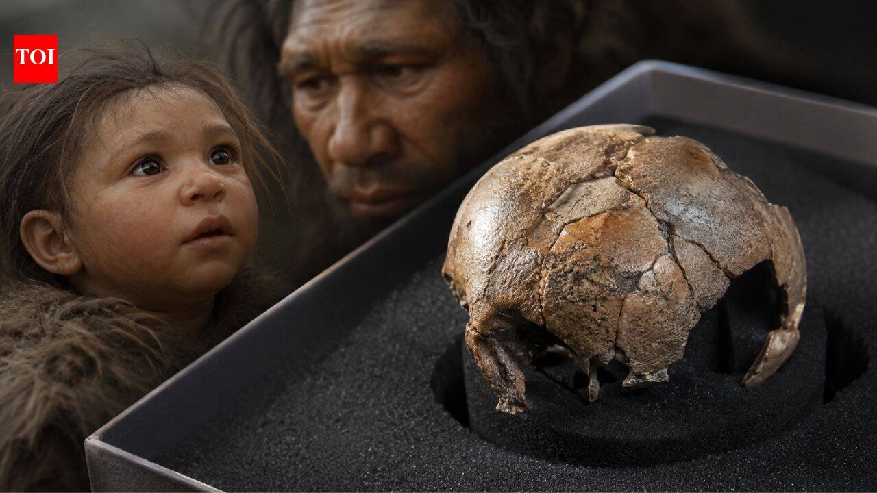
The first months of Neanderthal life remain one of the least understood chapters in human evolution. Fossils of adults and older children reveal the unique characteristics of these ancient relatives, but remains of unborn babies are extremely rare. This makes it difficult to determine when the familiar Neanderthal body shape actually began to emerge. A newly examined fossil from southern Germany is now helping to fill in some of the gaps. By studying the microstructure inside tiny fossil bones without damaging them, scientists have reconstructed the growth of a Neanderthal fetus in its final weeks before birth. Their results suggest that, at least before birth, Neanderthals followed a pattern of skeletal development that was strikingly similar to that of modern human infants, with many of their identifiable differences appearing only after they entered the world.
A cave in Germany preserves one of the earliest stages of Neanderthal life
The study was published in Royal Society Open Science and is titled “Early Neanderthal development revealed through virtual microanatomyThe fossil was revealed to have come from the Sesselfelsgrotte rock shelter in Bavaria, which houses one of the richest collections of Neanderthal remains in Europe, after decades of excavation. Although countless bones were discovered during excavations in the 1960s and 1970s, there are still tiny fragments that went unidentified for years because of their size.It was not until later investigations that experts realized that several of these delicate bones belonged to extremely young Neanderthals. Ancient DNA extracted from a lower leg bone has now confirmed that one of the men was an unborn Neanderthal, ending long-standing uncertainty about the identity of the remains.The specimen, known as Sesselfelsgrotte 1, is highly unusual. Fewer than ten Neanderthal fetuses, or newborns, have been discovered so far, making each discovery valuable for understanding how these ancient humans developed before birth.
See inside ancient bones without damaging them
The fetal skeleton survived except for a dozen tiny bone fragments, many of which were less than a few centimeters across. Because the material is so fragile, the team avoided traditional methods that would require cutting fossil bones.Instead, they rely on high-resolution imaging to create detailed three-dimensional views of the bones’ internal structures. This method allows scientists to examine microscopic growth patterns while keeping the fossil intact for future research, including further genetic analysis.Some fragments also showed faint surface marks consistent with carnivore digestion, suggesting that scavengers may have disturbed the remains at some point after death.
Fossil reveals striking similarities in fetal skeletal growth
Microscopic images show typical bone tissue of a rapidly developing fetus in late pregnancy. The blood-rich areas and newly formed bones are consistent with features commonly seen in modern human babies before birth.There are subtle differences between the various parts of the skeleton. The bones of the arms and legs appear to be slightly further back in development than the fragments of the skull and jaw. This change is not unexpected because different parts of the skeleton form in different ways.Long bones begin as cartilage and then gradually become mineralized, while many bones of the skull develop directly from connective tissue. The subtle changes seen in the fossils may simply reflect these individual biological pathways, rather than any unique Neanderthal patterns.The researchers say it’s too early to conclude that Neanderthal limbs always matured before the rest of the body because the evidence comes from a single fetus.
Teeth retain signs of early childhood health
In addition to the fetal skeleton, the scientists also examined two baby teeth from two different young Neanderthals from the same site.Inside each tooth, they identified areas where dentin (the hard tissue beneath tooth enamel) failed to fully mineralize during development. These small defects, called interglomerular dentin, can sometimes indicate an interruption in normal mineral formation as the tooth grows.This change may be related to periods of physiological stress, including vitamin D or calcium shortages, or problems affecting calcium absorption. Evidence cannot pinpoint the exact cause, and the disruption could occur at any point between late pregnancy and the first few years of life, when these teeth are forming.If confirmed, these defects would represent the earliest known evidence of such developmental disorders in Neanderthals.
The first few months of life made Neanderthals unique
Although unborn Neanderthals were very similar in skeletal development to modern human fetuses, these similarities did not persist throughout infancy.Previous research has shown that these pathways begin to diverge soon after birth. Modern human infants develop the rounded braincases familiar to us humans, while Neanderthal skulls retained a longer, lower profile. Scientists believe that most of this difference occurs in the first year of life, rather than before birth.The new findings fit neatly into this picture, suggesting that prenatal development follows a common blueprint before the two groups gradually take different developmental routes during infancy.